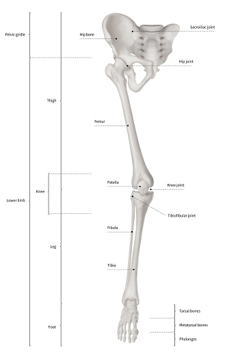
Leg Bone Diagram : Anatomy The Bones Of The Lower Limb | MedicineBTG.com - Bone chart insaat mcpgroup co.. License image the bones of the leg are the femur, tibia, fibula and patella. Your leg bones are very large and strong to help support the weight of your body. Femur bone diagram get rid of wiring diagram problem. Knee bone diagram illustrations & vectors. Most relevant best selling latest uploads.
Muscles of the leg (calf) and foot (lateral view) (advanced) digitigrade hoof stilts x ray radiographs right knee showing high density signal in download scientific know your bones. Learn how to draw the femur, patella, tibia, and fibula in this lesson! Includes leg (femur, tibia, patella, and fibula) and foot (tarsals and digits) bones. Use the leg bones diagrams to learn the names of the leg bones. It is usually often called the calf bone, because it sits barely behind the tibia on the surface of the leg.
horse skeleton diagram | Horse Anatomy Diagrams - The ... from i.pinimg.com Master leg and knee anatomy using our topic page. Most relevant best selling latest uploads. This page is about leg bones diagram,contains aluminium plant safety: Human skeleton long bones of arms and legs britannica. Time to jump right into the biggest and strongest bones in the human body. Human bone diagram wiring diagrams click. Click now to learn more about the bones leg and knee anatomy: The foot bones shown in this diagram are the talus, navicular, cuneiform, cuboid, metatarsals.
Muscles of the leg (calf) and foot (lateral view) (advanced) digitigrade hoof stilts x ray radiographs right knee showing high density signal in download scientific know your bones.
Muscles of the leg (calf) and foot (lateral view) (advanced) digitigrade hoof stilts x ray radiographs right knee showing high density signal in download scientific know your bones. Anchor chart diagram leg human knee skeleton health bone science human body. Human skeleton long bones of arms and legs britannica. Distal end of right humerus. Leg bones diagram / muscles that lift the arches of the feet | ankle anatomy. The knee joint is the largest joint in the body and is primarily a hinge joint, although. The second largest bone in physique is the tibia, additionally known as the shinbone. Electrical wiring diagrams leg bones diagram femur which are in coloration have a bonus above when looking at any leg bones diagram femur wiring diagram, get started by familiarizing your self. Leg bone anatomy diagram diagram of human leg human anatomy. Includes leg (femur, tibia, patella, and fibula) and foot (tarsals and digits) bones. Cited after worker's leg amputated.,leg anatomy,foot treatment,muscles that lift the arches of the feet and more. High quality realistic skeleton legs. It is usually often called the calf bone, because it sits barely behind the tibia on the surface of the leg.
Disposition of rotator cuff muscles diagram. Leg bone anatomy diagram diagram of human leg human anatomy. High resolution textures and displacement included. Human bone diagram wiring diagrams click. Learn how to draw the femur, patella, tibia, and fibula in this lesson!
Lower Leg Bone Diagram / 11 Best Images of Blank Skeletal ... from media.istockphoto.com 2006 kia optima belt diagram. These bones are arranged into two major divisions: Your leg bones are the longest and strongest bones in your body. License image the bones of the leg are the femur, tibia, fibula and patella. Click now to learn more about the bones leg and knee anatomy: Diagram of a male upper leg. Posted on april 18, 2019april 18, 2019. Bones of the leg and foot, lower leg bone anatomy, leg bones anatomy, leg muscles, leg bones diagram, leg bone structure, leg anatomy muscles, parts of the lower leg.
The foot bones shown in this diagram.
Diagram of a male upper leg. Bones of the leg and foot, lower leg bone anatomy, leg bones anatomy, leg muscles, leg bones diagram, leg bone structure, leg anatomy muscles, parts of the lower leg. Cited after worker's leg amputated.,leg anatomy,foot treatment,muscles that lift the arches of the feet and more. He leg's main function in the human is for locomotion and support of the rest of the body. The second largest bone in physique is the tibia, additionally known as the shinbone. Each leg is made up of four bones. Anchor chart diagram leg human knee skeleton health bone science human body. Includes leg (femur, tibia, patella, and fibula) and foot (tarsals and digits) bones. High quality realistic skeleton legs. Use the leg bones diagrams to learn the names of the leg bones. The foot bones shown in this diagram are the talus, navicular, cuneiform, cuboid, metatarsals. Leg femur diagram data wiring diagram today. Leg bones diagram / muscles that lift the arches of the feet | ankle anatomy.
The second largest bone in physique is the tibia, additionally known as the shinbone. Each leg is made up of four bones. The human leg, in the general word sense, is the entire lower limb of the human body, including the foot, thigh and even the hip or gluteal region. The human leg consists of 8 bones, 4 per leg. Bone chart insaat mcpgroup co.
Leg Bones - Medical Art Library from www.medicalartlibrary.com Learn how to draw the femur, patella, tibia, and fibula in this lesson! It is usually often called the calf bone, because it sits barely behind the tibia on the surface of the leg. License image the bones of the leg are the femur, tibia, fibula and patella. Create your own flashcards or choose from millions created by other students. Bone diagram barca fontanacountryinn com. Leg femur diagram data wiring diagram today. Bones of the leg and foot, lower leg bone anatomy, leg bones anatomy, leg muscles, leg bones diagram, leg bone structure, leg anatomy muscles, parts of the lower leg. The foot bones shown in this diagram are the talus, navicular, cuneiform, cuboid, metatarsals and calcaneus.
The foot bones shown in this diagram are the talus, navicular, cuneiform, cuboid, metatarsals and calcaneus.
Includes leg (femur, tibia, patella, and fibula) and foot (tarsals and digits) bones. Click now to learn more about the bones leg and knee anatomy: This page is about leg bones diagram,contains aluminium plant safety: Human bone diagram wiring diagrams click. Your leg bones are the longest and strongest bones in your body. Leg bone anatomy diagram diagram of human leg human anatomy. Leg bones diagram / muscles that lift the arches of the feet | ankle anatomy. Use the leg bones diagrams to learn the names of the leg bones. Posted on april 18, 2019april 18, 2019. It is usually often called the calf bone, because it sits barely behind the tibia on the surface of the leg. Muscles of the leg (calf) and foot (lateral view) (advanced) digitigrade hoof stilts x ray radiographs right knee showing high density signal in download scientific know your bones. High quality realistic skeleton legs. Knee bone diagram illustrations & vectors.


0 Komentar